
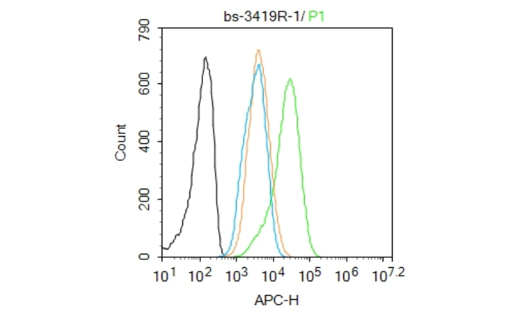
Blank control(black line):Hela.
Primary Antibody (green line): Rabbit Anti-Phospho-Smad2 (Ser465 + Ser467) antibody (bs-3419R)
Dilution:1ug/Test;
Secondary Antibody(white blue line): Goat anti-rabbit IgG-AF488
Dilution: 0.5ug/Test.
Isotype control(orange line): Normal Rabbit IgG
Protocol
The cells were fixed with 4% PFA (10min at room temperature)and then permeabilized with 90% ice-cold methanol for 20 min at -20℃, The cells were then incubated in 5%BSA to block non-specific protein-protein interactions for 30 min at room temperature .Cells stained with Primary Antibody for 30 min at room temperature. The secondary antibody used for 40 min at room temperature. Acquisition of 20,000 events was performed.
磷酸化细胞信号转导分子SMAD2抗体
Phospho-Smad2 (Ser465 + Ser467) Rabbit pAb
- bs-3419R
- 北京博奥森
- 北京市
- 现货
- 50ul
- 100ul
- 200ul
- 议价
- 2023-10-11 10:52:38
北京博奥森生物技术有限公司
一键申请试用
咨询
加入意向单
联系方式
- 英文名称
- Phospho-Smad2 (Ser465 + Ser467) Rabbit pAb
概述
产品编号
bs-3419R
产品分类
一抗
产品类型
磷酸化抗体、质检1级
英文名称
Phospho-Smad2 (Ser465 + Ser467) Rabbit pAb
中文名称
磷酸化细胞信号转导分子SMAD2抗体
英文别名
Smad2(Phospho S465 + S467); phospho-Smad2(p-Ser465/467); p-Smad2(Ser465/467); phospho-Smad2(p-S465/467); Smad2 (phospho S465 + S467); p-Smad2 (phospho S465 + S467); hMAD 2; hSMAD2; JV18 1; JV18; JV181; MAD; MAD Related Protein 2; MADH2; MADR2; MGC22139; MGC34440; Mothers Against Decapentaplegic Homolog 2; mothers against DPP homolog 2; SMAD 2; SMAD; SMAD2; SMAD2_HUMAN.
交叉反应
Human,Mouse,Rat(predicted:Chicken,Dog,Cow,Horse)
抗体来源
Rabbit
免疫原
KLH conjugated Synthesised phosphopeptide derived from human SMAD2 around the phosphorylation site of Ser465/467
亚型
IgG
纯化方法
affinity purified by Protein A
克隆类型
Polyclonal
理论分子量
58kDa
浓度
1mg/ml
储存液
0.01M TBS(pH7.4) with 1% BSA, 0.03% Proclin300 and 50% Glycerol.
保存条件
Shipped at 4℃. Store at -20 °C for one year. Avoid repeated freeze/thaw cycles.
Subunit
Momomer; the absence of TGF-beta. Heterodimer; in the presence of TGF-beta. Forms a heterodimer with co-SMAD, SMAD4, in the nucleus to form the transactivation complex SMAD2/SMAD4. Interacts with AIP1, HGS, PML and WWP1. Interacts with NEDD4L in response to TGF-beta. Found in a complex with SMAD3 and TRIM33 upon addition of TGF-beta. Interacts with ACVR1B, SMAD3 and TRIM33. Interacts (via the MH2 domain) with ZFYVE9; may form trimers with the SMAD4 co-SMAD. Interacts with FOXH1, homeobox protein TGIF, PEBP2-alpha subunit, CREB-binding protein (CBP), EP300 and SKI. Interacts with SNON; when phosphorylated at Ser-465/467. Interacts with SKOR1 and SKOR2. Interacts with PRDM16. Interacts (via MH2 domain) with LEMD3. Interacts with RBPMS. Interacts with WWP1. Interacts (dephosphorylated form, via the MH1 and MH2 domains) with RANBP3 (via its C-terminal R domain); the interaction results in the export of dephosphorylated SMAD3 out of the nucleus and termination ot the TGF-beta signaling. Interacts with PDPK1 (via PH domain).
Subcellular Location
Cytoplasm. Nucleus. Note=Cytoplasmic and nuclear in the absence of TGF-beta. On TGF-beta stimulation, migrates to the nucleus when complexed with SMAD4. On dephosphorylation by phosphatase PPM1A, released from the SMAD2/SMAD4 complex, and exported out of the nucleus by interaction with RANBP1.
Tissue Specificity
Expressed at high levels in skeletal muscle, heart and placenta.
Post-translational modifications
Phosphorylated on one or several of Thr-220, Ser-245, Ser-250, and Ser-255. In response to TGF-beta, phosphorylated on Ser-465/467 by TGF-beta and activin type 1 receptor kinases. Able to interact with SMURF2 when phosphorylated on Ser-465/467, recruiting other proteins, such as SNON, for degradation. In response to decorin, the naturally occurring inhibitor of TGF-beta signaling, phosphorylated on Ser-240 by CaMK2. Phosphorylated by MAPK3 upon EGF stimulation; which increases transcriptional activity and stability, and is blocked by calmodulin. Phosphorylated by PDPK1.
In response to TGF-beta, ubiquitinated by NEDD4L; which promotes its degradation.
Acetylated on Lys-19 by coactivators in response to TGF-beta signaling, which increases transcriptional activity. Isoform short: Acetylation increases DNA binding activity in vitro and enhances its association with target promoters in vivo. Acetylation in the nucleus by EP300 is enhanced by TGF-beta.
Similarity
Belongs to the dwarfin/SMAD family.
Contains 1 MH1 (MAD homology 1) domain.
Contains 1 MH2 (MAD homology 2) domain.
Contains 1 MH1 (MAD homology 1) domain.
Contains 1 MH2 (MAD homology 2) domain.
Database links
Entrez Gene: 4087 Human
Entrez Gene: 17126 Mouse
Omim: 601366 Human
SwissProt: Q15796 Human
SwissProt: Q62432 Mouse
Unigene: 12253 Human
Unigene: 705764 Human
Unigene: 391091 Mouse
Unigene: 2755 Rat
背景资料
The protein encoded by this gene belongs to the SMAD, a family of proteins similar to the gene products of the Drosophila gene 'mothers against decapentaplegic' (Mad) and the C. elegans gene Sma. SMAD proteins are signal transducers and transcriptional modulators that mediate multiple signaling pathways. This protein mediates the signal of the transforming growth factor (TGF)-beta, and thus regulates multiple cellular processes, such as cell proliferation, apoptosis, and differentiation. This protein is recruited to the TGF-beta receptors through its interaction with the SMAD anchor for receptor activation (SARA) protein. In response to TGF-beta signal, this protein is phosphorylated by the TGF-beta receptors. The phosphorylation induces the dissociation of this protein with SARA and the association with the family member SMAD4. The association with SMAD4 is important for the translocation of this protein into the nucleus, where it binds to target promoters and forms a transcription repressor complex with other cofactors. This protein can also be phosphorylated by activin type 1 receptor kinase, and mediates the signal from the activin. Alternatively spliced transcript variants have been observed for this gene. [provided by RefSeq, May 2012]
标记抗体
应用
| 应用 | 推荐稀释比例 |
|---|---|
| ELISA | 1:5000-10000 |
| Flow-Cyt | 1ug/Test |
| IF | 1:100-500 |
| ICC | 1:100 |
| IHC-F | 1:100-500 |
| IHC-P | 1:100-500 |
| WB | 1:500-2000 |
图片资料
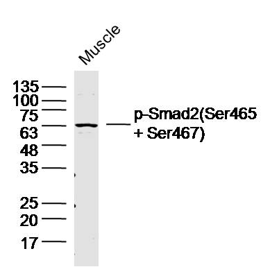
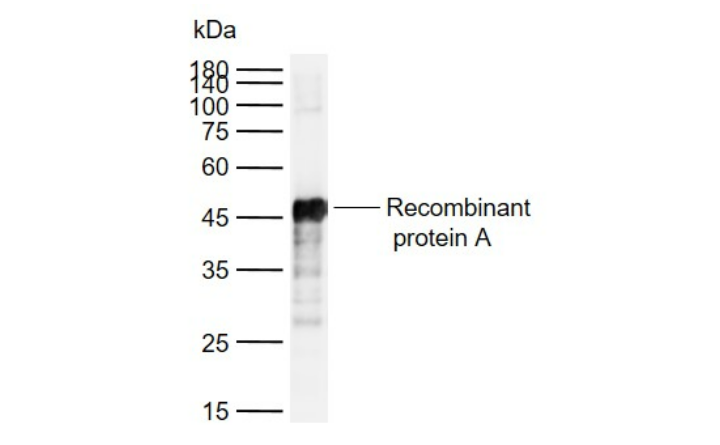
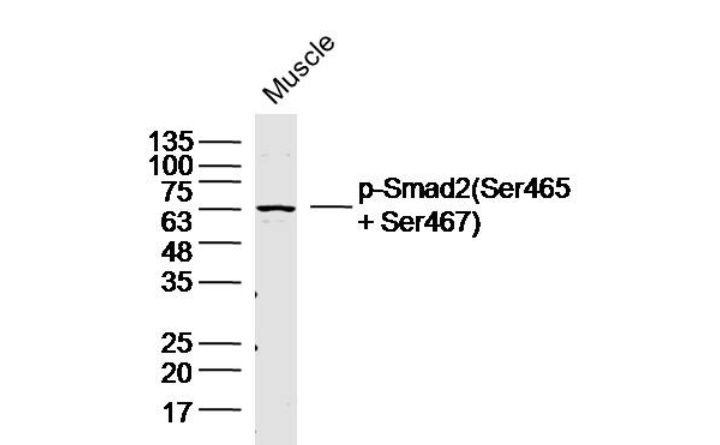
Sample:Muscle (Mouse)Lysate at 40 ug
Primary: Anti-p-Smad2(Ser465+Ser467)(bs-3419R)at 1/300 dilution
Secondary: IRDye800CW Goat Anti-RabbitIgG at 1/20000 dilution
Predicted band size: 58kD
Observed band size: 63kD
Primary: Anti-p-Smad2(Ser465+Ser467)(bs-3419R)at 1/300 dilution
Secondary: IRDye800CW Goat Anti-RabbitIgG at 1/20000 dilution
Predicted band size: 58kD
Observed band size: 63kD
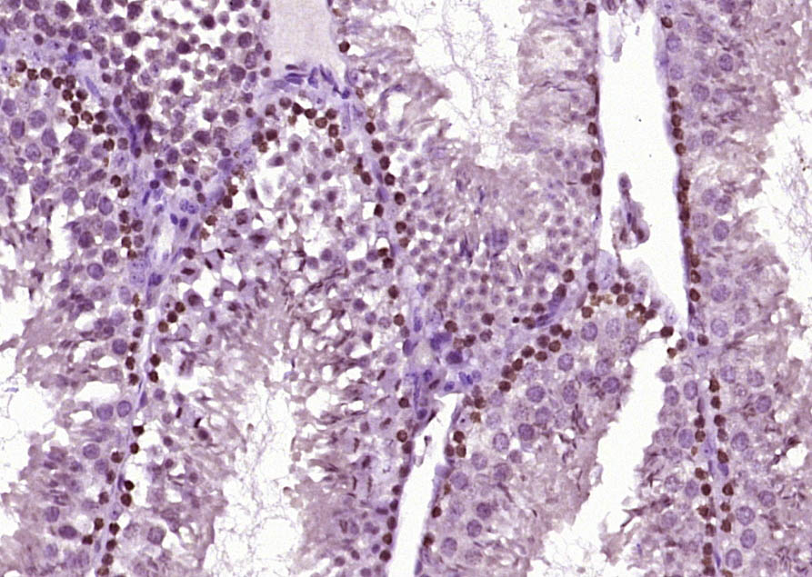
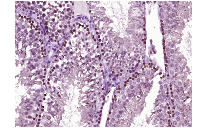
Paraformaldehyde-fixed, paraffin embedded (Mouse testis); Antigen retrieval by boiling in sodium citrate buffer (pH6.0) for 15min; Block endogenous peroxidase by 3% hydrogen peroxide for 20 minutes; Blocking buffer (normal goat serum) at 37°C for 30min; Antibody incubation with (Phospho-Smad2(Ser465 + Ser467)) Polyclonal Antibody, Unconjugated (bs-3419R) at 1:400 overnight at 4°C, followed by operating according to SP Kit(Rabbit) (sp-0023) instructionsand DAB staining.
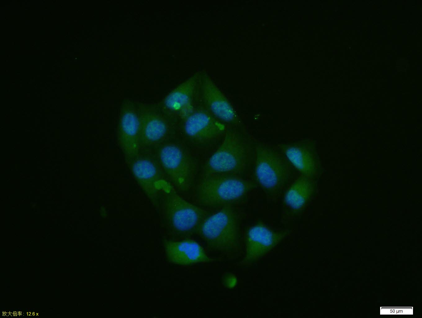
Hela cell; 4% Paraformaldehyde-fixed; Triton X-100 at room temperature for 20 min; Blocking buffer (normal goat serum, C-0005) at 37°C for 20 min; Antibody incubation with (Phospho-Smad2 (Ser465 + Ser467) ) polyclonal Antibody, Unconjugated (bs-3419R) 1:100, 90 minutes at 37°C; followed by a conjugated Goat Anti-Rabbit IgG antibody at 37°C for 90 minutes, DAPI (blue, C02-04002) was used to stain the cell nuclei.
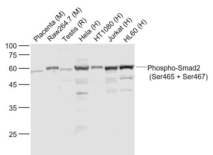
Sample:
Lane 1: Placenta (Mouse) Lysate at 40 ug
Lane 2: Raw264.7 (Mouse) Cell Lysate at 30 ug
Lane 3: Testis (Rat) Lysate at 40 ug
Lane 4: Hela (Human) Cell Lysate at 30 ug
Lane 5: HT1080 (Human) Cell Lysate at 30 ug
Lane 6: Jurkat (Human) Cell Lysate at 30 ug
Lane 7: HL60 (Human) Cell Lysate at 30 ug
Primary: Anti-Phospho-Smad2 (Ser465 + Ser467) (bs-3419R) at 1/1000 dilution
Secondary: IRDye800CW Goat Anti-Rabbit IgG at 1/20000 dilution
Predicted band size: 60 kD
Observed band size: 60 kD
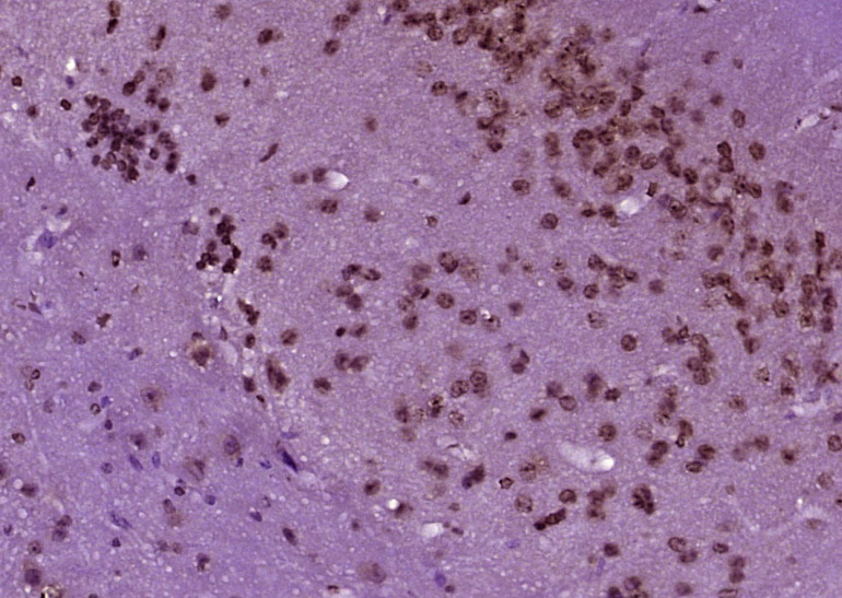
Paraformaldehyde-fixed, paraffin embedded (Mouse brain); Antigen retrieval by boiling in sodium citrate buffer (pH6.0) for 15min; Block endogenous peroxidase by 3% hydrogen peroxide for 20 minutes; Blocking buffer (normal goat serum) at 37°C for 30min; Antibody incubation with (Phospho-Smad2(Ser465 + Ser467)) Polyclonal Antibody, Unconjugated (bs-3419R) at 1:400 overnight at 4°C, followed by operating according to SP Kit(Rabbit) (sp-0023) instructionsand DAB staining.