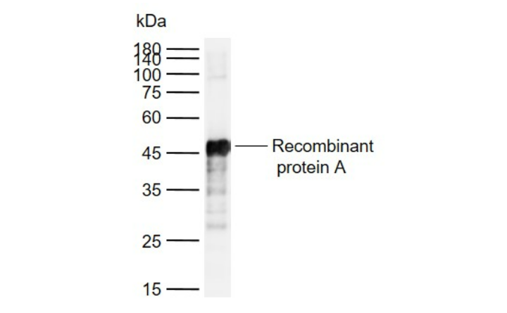

Sample:
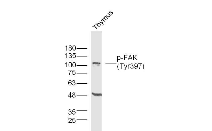
Thymus (Mouse) Lysate at 40 ug
Primary: Anti- p-FAK (Tyr397) (bs-3159R)at 1/300 dilution
Secondary: IRDye800CW Goat Anti-Rabbit IgG at 1/20000 dilution
Predicted band size: 116 kD
Observed band size: 116 kD
磷酸化粘着斑激酶抗体
Phospho-FAK (Tyr397) Rabbit pAb
- bs-3159R
- 北京博奥森
- 北京市
- 现货
- 按需
- 议价
- 2023-10-19 15:02:40
北京博奥森生物技术有限公司
一键申请试用
咨询
加入意向单
联系方式
- 英文名称
- Phospho-FAK (Tyr397) Rabbit pAb
概述
产品编号
bs-3159R
产品分类
一抗
产品类型
磷酸化抗体、质检1级
英文名称
Phospho-FAK (Tyr397) Rabbit pAb
中文名称
磷酸化粘着斑激酶抗体
英文别名
FAK (phospho Y397); p-FAK (phospho Y397); PTK2(phospho Y397); p-PTK2; FADK 1; FADK; FAK 1; FAK related non kinase polypeptide; FAK1; Focal adhesion kinase 1; FRNK; pp125FAK; Protein tyrosine kinase 2; Protein Tyrosine Kinase Cytoplasmic; PTK 2; FAK1_HUMAN; Focal adhesion kinase-related nonkinase; Protein phosphatase 1 regulatory subunit 71; PPP1R71; Protein-tyrosine kinase 2; p125FAK.
产品应用
WB, IHC-P, IHC-F, IF, ELISA
交叉反应
Human,Mouse,Rat(predicted:Chicken,Dog,Cow,Horse,Rabbit)
抗体来源
Rabbit
免疫原
KLH cunjugated Synthesised phosphopeptide derived from human FAK around the phosphorylation site of Tyr397:DD(p-Y)AE
亚型
IgG
纯化方法
affinity purified by Protein A
克隆类型
Polyclonal
理论分子量
116kDa
浓度
1mg/ml
储存液
0.01M TBS(pH7.4) with 1% BSA, 0.03% Proclin300 and 50% Glycerol.
保存条件
Shipped at 4℃. Store at -20 °C for one year. Avoid repeated freeze/thaw cycles.
功能
Non-receptor protein-tyrosine kinase that plays an essential role in regulating cell migration, adhesion, spreading, reorganization of the actin cytoskeleton, formation and disassembly of focal adhesions and cell protrusions, cell cycle progression, cell proliferation and apoptosis. Required for early embryonic development and placenta development. Required for embryonic angiogenesis, normal cardiomyocyte migration and proliferation, and normal heart development. Regulates axon growth and neuronal cell migration, axon branching and synapse formation; required for normal development of the nervous system. Plays a role in osteogenesis and differentiation of osteoblasts. Functions in integrin signal transduction, but also in signaling downstream of numerous growth factor receptors, G-protein coupled receptors (GPCR), EPHA2, netrin receptors and LDL receptors. Forms multisubunit signaling complexes with SRC and SRC family members upon activation; this leads to the phosphorylation of additional tyrosine residues, creating binding sites for scaffold proteins, effectors and substrates. Regulates numerous signaling pathways. Promotes activation of phosphatidylinositol 3-kinase and the AKT1 signaling cascade. Promotes activation of MAPK1/ERK2, MAPK3/ERK1 and the MAP kinase signaling cascade. Promotes localized and transient activation of guanine nucleotide exchange factors (GEFs) and GTPase-activating proteins (GAPs), and thereby modulates the activity of Rho family GTPases. Signaling via CAS family members mediates activation of RAC1. Recruits the ubiquitin ligase MDM2 to P53/TP53 in the nucleus, and thereby regulates P53/TP53 activity, P53/TP53 ubiquitination and proteasomal degradation. Phosphorylates SRC; this increases SRC kinase activity. Phosphorylates ACTN1, ARHGEF7, GRB7, RET and WASL. Promotes phosphorylation of PXN and STAT1; most likely PXN and STAT1 are phosphorylated by a SRC family kinase that is recruited to autophosphorylated PTK2/FAK1, rather than by PTK2/FAK1 itself. Promotes phosphorylation of BCAR1; GIT2 and SHC1; this requires both SRC and PTK2/FAK1. Promotes phosphorylation of BMX and PIK3R1. Isoform 6 (FRNK) does not contain a kinase domain and inhibits PTK2/FAK1 phosphorylation and signaling. Its enhanced expression can attenuate the nuclear accumulation of LPXN and limit its ability to enhance serum response factor (SRF)-dependent gene transcription.
亚基
Interacts (via first Pro-rich region) with CAS family members (via SH3 domain), including BCAR1, BCAR3, CASS4 and NEDD9. Interacts with GIT1. Interacts with SORBS1. Interacts with RGNEF. Interacts with SHB. Interacts with PXN and TLN1. Interacts with STAT1. Interacts with DCC. Interacts with WASL. Interacts with ARHGEF7. Interacts with GRB2 and GRB7 (By similarity). Component of a complex that contains at least FER, CTTN and PTK2/FAK1. Interacts with BMX. Interacts with TGFB1I1. Interacts with STEAP4. Interacts with ZFYVE21. Interacts with ESR1. Interacts with PIK3R1 or PIK3R2. Interacts with SRC, FGR, FLT4 and RET. Interacts with EPHA2 in resting cells; activation of EPHA2 recruits PTPN11, leading to dephosphorylation of PTK2/FAK1 and dissociation of the complex. Interacts with EPHA1 (kinase activity-dependent). Interacts with CD4; this interaction requires the presence of HIV-1 gp120. Interacts with PIAS1. Interacts with ARHGAP26 and SHC1. Interacts with RB1CC1; this inhibits PTK2/FAK1 activity and activation of downstream signaling pathways. Interacts with P53/TP53 and MDM2. Interacts with LPXN (via LD motif 3).
亚细胞定位
Cell junction, focal adhesion. Cell membrane; Peripheral membrane protein; Cytoplasmic side. Cytoplasm, cell cortex. Cytoplasm, cytoskeleton. Cytoplasm, cytoskeleton, centrosome. Nucleus. Note=Constituent of focal adhesions. Detected at microtubules.
组织特异性
Detected in B and T-lymphocytes. Isoform 1 and isoform 6 are detected in lung fibroblasts (at protein level). Ubiquitous.
翻译后修饰
Phosphorylated on tyrosine residues upon activation, e.g. upon integrin signaling. Tyr-397 is the major autophosphorylation site, but other kinases can also phosphorylate this residue. Phosphorylation at Tyr-397 promotes interaction with SRC and SRC family members, leading to phosphorylation at Tyr-576, Tyr-577 and at additional tyrosine residues. FGR promotes phosphorylation at Tyr-397 and Tyr-576. FER promotes phosphorylation at Tyr-577, Tyr-861 and Tyr-925, even when cells are not adherent. Tyr-397, Tyr-576 and Ser-722 are phosphorylated only when cells are adherent. Phosphorylation at Tyr-397 is important for interaction with BMX, PIK3R1 and SHC1. Phosphorylation at Tyr-925 is important for interaction with GRB2. Dephosphorylated by PTPN11; PTPN11 is recruited to PTK2 via EPHA2 (tyrosine phosphorylated). Microtubule-induced dephosphorylation at Tyr-397 is crucial for the induction of focal adhesion disassembly; this dephosphorylation could be catalyzed by PTPN11 and regulated by ZFYVE21.
Sumoylated; this enhances autophosphorylation.
Sumoylated; this enhances autophosphorylation.
疾病
Note=Aberrant PTK2/FAK1 expression may play a role in cancer cell proliferation, migration and invasion, in tumor formation and metastasis. PTK2/FAK1 overexpression is seen in many types of cancer.
相似性
Belongs to the protein kinase superfamily. Tyr protein kinase family.
FAK subfamily.
Contains 1 FERM domain.
FAK subfamily.
Contains 1 FERM domain.
数据库链接
Entrez Gene: 5747 Human
Entrez Gene: 14083 Mouse
Omim: 600758 Human
SwissProt: Q05397 Human
SwissProt: P34152 Mouse
Unigene: 395482 Human
Unigene: 254494 Mouse
Unigene: 2809 Rat
背景资料
Non-receptor protein-tyrosine kinase implicated in signaling pathways involved in cell motility, proliferation and apoptosis. Activated by tyrosine-phosphorylation in response to either integrin clustering induced by cell adhesion or antibody cross-linking, or via G-protein coupled receptor (GPCR) occupancy by ligands such as bombesin or lysophosphatidic acid, or via LDL receptor occupancy. Plays a potential role in oncogenic transformations resulting in increased kinase activity. [SUBCELLULAR LOCATION] Cell junction, focal adhesion. Cell membrane; Peripheral membrane protein; Cytoplasmic side. Note=Constituent of focal adhesions.
应用
| 应用 | 推荐稀释比例 |
|---|---|
| ELISA | 1:5000-10000 |
| IF | 1:100-500 |
| IHC-F | 1:100-500 |
| IHC-P | 1:100-500 |
| WB | 1:500-2000 |
图片资料

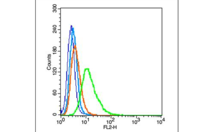
Blank control (blue line): A549 (fixed with 2% paraformaldehyde (10 min) , then permeabilized with 90% ice-cold methanol for 30 min on ice).
Primary Antibody (green line): Rabbit Anti-Phospho-FAK (Tyr397) antibody (bs-3159R),Dilution: 1μg/10^6 cells.
Isotype Control Antibody (orange line): Rabbit IgG.
Secondary Antibody (white blue line): Goat anti-rabbit IgG-PE,Dilution: 1μg /test.
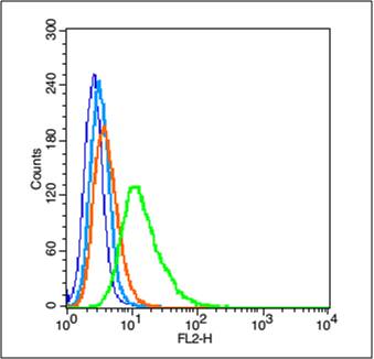
Primary Antibody (green line): Rabbit Anti-Phospho-FAK (Tyr397) antibody (bs-3159R),Dilution: 1μg/10^6 cells.
Isotype Control Antibody (orange line): Rabbit IgG.
Secondary Antibody (white blue line): Goat anti-rabbit IgG-PE,Dilution: 1μg /test.
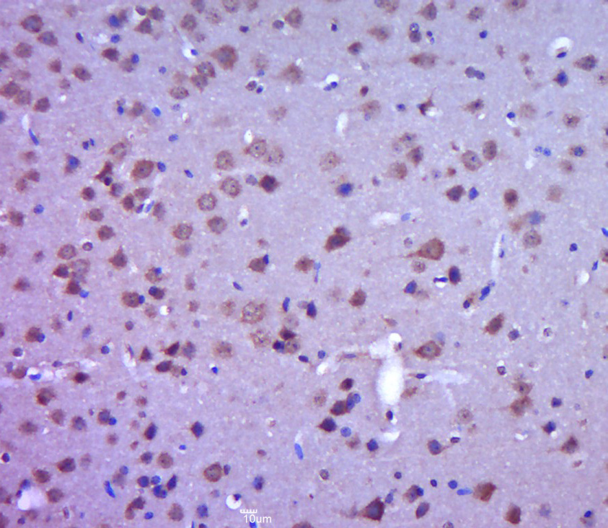
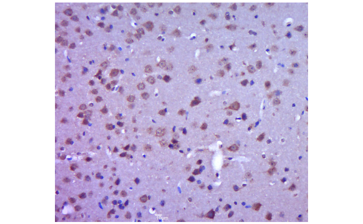
Paraformaldehyde-fixed, paraffin embedded (mouse brain tissue); Antigen retrieval by boiling in sodium citrate buffer (pH6.0) for 15min; Block endogenous peroxidase by 3% hydrogen peroxide for 20 minutes; Blocking buffer (normal goat serum) at 37°C for 30min; Antibody incubation with (P-FAK (Tyr397)) Polyclonal Antibody, Unconjugated (bs-3159R) at 1:400 overnight at 4°C, followed by a conjugated secondary (sp-0023) for 20 minutes and DAB staining.
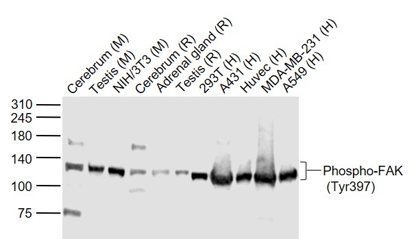
Sample:
Lane 1: Cerebrum (Mouse) Lysate at 40 ug
Lane 2: Testis (Mouse) Lysate at 40 ug
Lane 3: NIH/3T3 (Mouse) Cell Lysate at 30 ug
Lane 4: Cerebrum (Rat) Lysate at 40 ug
Lane 5: Adrenal gland (Rat) Lysate at 40 ug
Lane 6: Testis (Rat) Lysate at 40 ug
Lane 7: 293T (Human) Cell Lysate at 30 ug
Lane 8: A431 (Human) Cell Lysate at 30 ug
Lane 9: Huvec (Human) Cell Lysate at 30 ug
Lane 10: MDA-MB-231 (Human) Cell Lysate at 30 ug
Lane 11: A549 (Human) Cell Lysate at 30 ug
Primary: Anti-Phospho-FAK (Tyr397) (bs-3159R) at 1/1000 dilution
Secondary: IRDye800CW Goat Anti-Rabbit IgG at 1/20000 dilution
Predicted band size: 125 kD
Observed band size: 125 kD
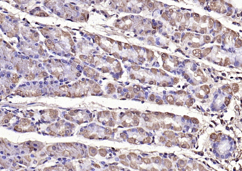
Paraformaldehyde-fixed, paraffin embedded (human gastric carcinoma); Antigen retrieval by boiling in sodium citrate buffer (pH6.0) for 15min; Block endogenous peroxidase by 3% hydrogen peroxide for 20 minutes; Blocking buffer (normal goat serum) at 37°C for 30min; Antibody incubation with (Phospho-FAK (Tyr397)) Polyclonal Antibody, Unconjugated (bs-3159R) at 1:200 overnight at 4°C, followed by operating according to SP Kit(Rabbit) (sp-0023) instructionsand DAB staining.