
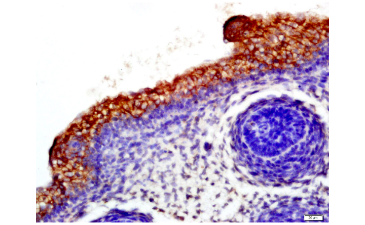
Tissue/cell: mouse fetal skin; 4% Paraformaldehyde-fixed and paraffin-embedded;
Antigen retrieval: citrate buffer ( 0.01M, pH 6.0 ), Boiling bathing for 15min; Block endogenous peroxidase by 3% Hydrogen peroxide for 30min; Blocking buffer (normal goat serum,C-0005) at 37℃ for 20 min;
Incubation: Anti-CD19 Polyclonal Antibody, Unconjugated(bs-0079R) 1:500, overnight at 4°C, followed by conjugation to the secondary antibody(SP-0023) and DAB(C-0010) staining

CD19抗体
CD19 Rabbit pAb
- bs-0079R
- 北京博奥森
- 北京市
- 现货
- 50ul
- 100ul
- 200ul
- 议价
- 2023-10-18 13:52:13
北京博奥森生物技术有限公司
一键申请试用
咨询
加入意向单
联系方式
- 英文名称
- CD19 Rabbit pAb
概述
产品编号
bs-0079R
产品分类
一抗
英文名称
CD19 Rabbit pAb
中文名称
CD19抗体
英文别名
CD19_HUMAN; B-lymphocyte antigen CD19; B-lymphocyte surface antigen B4; Differentiation antigen CD19; T-cell surface antigen Leu-12; CD_antigen: CD19; B-lymphocyte surface antigen B4; Leu 12; Leu12;
交叉反应
Human,Mouse,Rat(predicted:Pig,Cow,Horse,GuineaPig)
抗体来源
Rabbit
免疫原
KLH conjugated synthetic peptide derived from human CD19
亚型
IgG
纯化方法
affinity purified by Protein A
克隆类型
Polyclonal
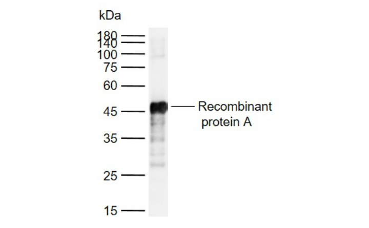
理论分子量
59kDa
检测分子量
95 kDa
浓度
1mg/ml
储存液
0.01M TBS(pH7.4) with 1% BSA, 0.03% Proclin300 and 50% Glycerol.
保存条件
Shipped at 4℃. Store at -20 °C for one year. Avoid repeated freeze/thaw cycles.
功能
Assembles with the antigen receptor of B-lymphocytes in order to decrease the threshold for antigen receptor-dependent stimulation.
亚基
Forms a complex with CD21, CD81 and CD225 in the membrane of mature B-cells. Interacts with VAV. Interacts with GRB2 and SOS when phosphorylated on Tyr-348 and/or Tyr-378. Interacts with PLCG2 when phosphorylated on Tyr-409. Interacts with LYN.
亚细胞定位
Membrane; Single-pass type I membrane protein.
翻译后修饰
Phosphorylated on serine and threonine upon DNA damage, probably by ATM or ATR. Phosphorylated on tyrosine following B-cell activation. Phosphorylated on tyrosine residues by LYN.
疾病
Defects in CD19 are the cause of immunodeficiency common variable type 3 (CVID3) [MIM:613493]; also called antibody deficiency due to CD19 defect. CVID3 is a primary immunodeficiency characterized by antibody deficiency, hypogammaglobulinemia, recurrent bacterial infections and an inability to mount an antibody response to antigen. The defect results from a failure of B-cell differentiation and impaired secretion of immunoglobulins; the numbers of circulating B-cells is usually in the normal range, but can be low.
相似性
Belongs to the selectin/LECAM family.
Contains 2 Ig-like C2-type (immunoglobulin-like) domains.
Contains 2 Ig-like C2-type (immunoglobulin-like) domains.
数据库链接
Entrez Gene: 930 Human
Entrez Gene: 12478 Mouse
Omim: 107265 Human
SwissProt: P15391 Human
SwissProt: P25918 Mouse
Unigene: 652262 Human
Unigene: 4360 Mouse
背景资料
This gene encodes a member of the immunoglobulin gene superfamily. Expression of this cell surface protein is restricted to B cell lymphocytes. This protein is a reliable marker for pre-B cells but its expression diminishes during terminal B cell differentiation in antibody secreting plasma cells. The protein has two N-terminal extracellular Ig-like domains separated by a non-Ig-like domain, a hydrophobic transmembrane domain, and a large C-terminal cytoplasmic domain. This protein forms a complex with several membrane proteins including complement receptor type 2 (CD21) and tetraspanin (CD81) and this complex reduces the threshold for antigen-initiated B cell activation. Activation of this B-cell antigen receptor complex activates the phosphatidylinositol 3-kinase signalling pathway and the subsequent release of intracellular stores of calcium ions. This protein is a target of chimeric antigen receptor (CAR) T-cells used in the treatment of lymphoblastic leukemia. Mutations in this gene are associated with the disease common variable immunodeficiency 3 (CVID3) which results in a failure of B-cell differentiation and impaired secretion of immunoglobulins. CVID3 is characterized by hypogammaglobulinemia, an inability to mount an antibody response to antigen, and recurrent bacterial infections. Alternative splicing results in multiple transcript variants encoding distinct isoforms. [provided by RefSeq, Jul 2020]
应用
| 应用 | 推荐稀释比例 |
|---|---|
| ELISA | 1:5000-10000 |
| Flow-Cyt | 1μg/Test |
| IF | 1:100-500 |
| IHC-F | 1:100-500 |
| IHC-P | 1:100-500 |
图片资料

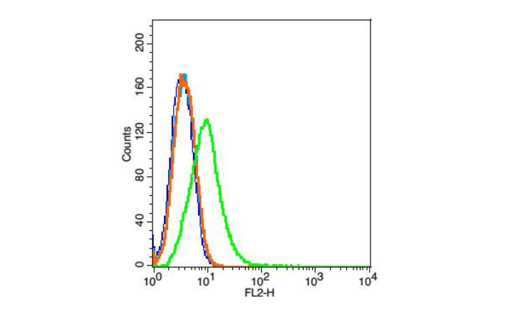
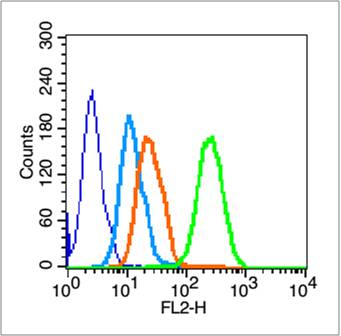
Blank control: Raji(blue).
Primary Antibody: Rabbit Anti-CD19 antibody(bs-0079R), Dilution: 5μg in 100 μL 1X PBS containing 0.5% BSA;
Isotype Control Antibody: Rabbit IgG (orange) ,used under the same conditions.
Secondary Antibody: Goat anti-rabbit IgG-PE(white blue), Dilution: 1:200 in 1 X PBS containing 0.5% BSA.
Protocol
Primary antibody (bs-0079R, 5μg /1x10^6 cells) were incubated for 30 min on the ice, followed by 1 X PBS containing 0.5% BSA + 1 0% goat serum (15 min) to block non-specific protein-protein interactions. Then the Goat Anti-rabbit IgG/PE antibody was added into the blocking buffer mentioned above to react with the primary antibody at 1/200 dilution for 30 min on ice. Acquisition of 20,000 events was performed.
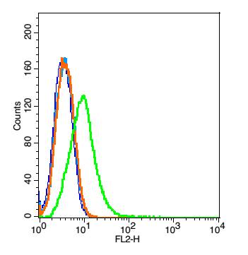
Blank control (blue line): HL60 cells (blue).
Primary Antibody (green line): Rabbit Anti-CD19 antibody (bs-0079R)
Dilution: 1μg /10^6 cells;
Isotype Control Antibody (orange line): Rabbit IgG .
Secondary Antibody (white blue line): Goat anti-rabbit IgG-PE
Dilution: 1μg /test.
Protocol
The cells were fixed with 70% methanol (Overnight at 4℃) . Cells stained with Primary Antibody for 30 min at room temperature. The cells were then incubated in 1 X PBS/2%BSA/10% goat serum to block non-specific protein-protein interactions followed by the antibody for 15 min at room temperature. The secondary antibody used for 40 min at room temperature. Acquisition of 20,000 events was performed.
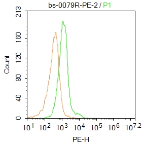
Blank control: Mouse spleen.
Primary Antibody (green line): Rabbit Anti-CD19 antibody (bs-0079R-PE)
Dilution: 2μg /10^6 cells;
Isotype Control Antibody (orange line): Rabbit IgG .
Protocol
The cells were incubated in 5%BSA to block non-specific protein-protein interactions for 30 min at at room temperature .Cells stained with Primary Antibody for 30 min at room temperature. Acquisition of 20,000 events was performed.