
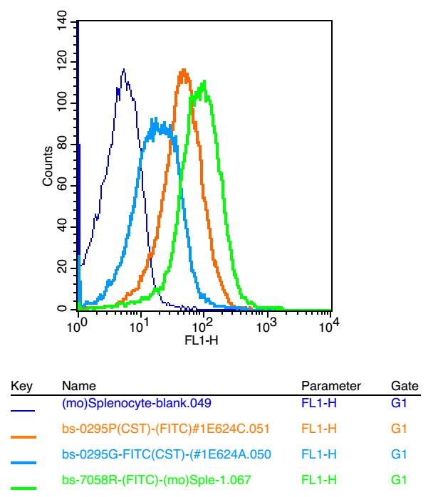
Blank control:A431.
Primary Antibody (green line): Rabbit Anti-ADGRE1 antibody (bs-7058R)
Dilution: 2ug/Test;
Secondary Antibody : Goat anti-rabbit IgG-AF488
Dilution: 0.5ug/Test.
Protocol
The cells were incubated in 5%BSA to block non-specific protein-protein interactions for 30 min at room temperature .Cells stained with Primary Antibody for 30 min at room temperature. The secondary antibody used for 40 min at room temperature. Acquisition of 20,000 events was performed.

FITC标记的表皮生长因子样激素受体1(EMR1)抗体
ADGRE1/FITC
- bs-7058R-FITC
- 北京博奥森
- 北京市
- 现货
- 100ul
- 议价
- 2023-10-11 14:40:22
北京博奥森生物技术有限公司
一键申请试用
咨询
加入意向单
联系方式
- 英文名称
- ADGRE1/FITC
概述
产品编号
bs-7058R-FITC
产品分类
标记一抗
英文名称
ADGRE1/FITC
中文名称
FITC标记的表皮生长因子样激素受体1(EMR1)抗体
英文别名
F4/80; Adhesion G protein-coupled receptor E1; Cell surface glycoprotein EMR1; Cell surface glycoprotein F4/80; DD7A5 7; Egf like module containing mucin like hormone receptor like 1; Egf like module containing mucin like hormone receptor like sequence 1; EGF like module receptor 1; EGF TM7; EGF-like module receptor 1; EGF-like module-containing mucin-like hormone receptor-like 1; EGFTM7; EMR 1; EMR1; EMR-1; EMR1 hormone receptor; EMR1_HUMAN; AGRE1_HUMAN; Gpf480; Ly71; Lymphocyte antigen 71; TM7LN3.
交叉反应
Human,Mouse(predicted:Rat,Pig,GuineaPig)
标记
FITC
抗体来源
Rabbit
免疫原
KLH conjugated synthetic peptide derived from human ADGRE1
亚型
IgG
纯化方法
affinity purified by Protein A
克隆类型
Polyclonal
浓度
1mg/ml
储存液
0.01M TBS(pH7.4) with 1% BSA, 0.03% Proclin300 and 50% Glycerol.
保存条件
Shipped at 4℃. Store at -20 °C for one year. Avoid repeated freeze/thaw cycles.
Subunit
Belongs to the G-protein coupled receptor 2 family. LN-TM7 subfamily. Contains 6 EGF-like domains. Contains 1 GPS domain.
Subcellular Location
Cell membrane.
Tissue Specificity
Wide expression; increased levels in peripheral blood mononuclear cells.
Similarity
Belongs to the G-protein coupled receptor 2 family. LN-TM7 subfamily.
Contains 6 EGF-like domains.
Contains 1 GPS domain.
Contains 6 EGF-like domains.
Contains 1 GPS domain.
Database links
Entrez Gene: 2015 Human
Entrez Gene: 13733 Mouse
Omim: 600493 Human
SwissProt: Q14246 Human
SwissProt: Q61549 Mouse
Unigene: 2375 Human
Unigene: 2254 Mouse
背景资料
The epidermal growth factor (EGF)-TM7 family constitutes a group of class B G-protein coupled receptors, which includes CD97, EMR1 (EGF-like molecule containing mucin-like hormone receptor 1, designated F4/80 in mouse), EMR2, EMR3, FIRE, and ETL (1–3). These family members are characterized by an extended extracellular region with several N-terminal EGF domains, and are predominantly expressed on cells of the immune system (1–3). The EGF-TM7 protein family are encoded by a gene cluster on human chromosome 19p13 (1,3,4). The F4/80 molecule is solely expressed on the surface of macrophages and serves as a marker for mature macrophage tissues, including Kupffer cells in liver, splenic red pulp macrophages, brain microglia, gut lamina propria, and Langerhans cells in the skin (1). F4/80/EMR1 undergoes extensive N-linked glycosylation as well as some O-linked glycosylation (5,6). The function of F4/80/EMR1 is unclear, but it is speculated to be involved in macrophage adhesion events, cell migration, or as a G-protein coupled signaling component of macrophages.
应用
| 应用 | 推荐稀释比例 |
|---|---|
| Flow-Cyt | 2μg/Test |
图片资料

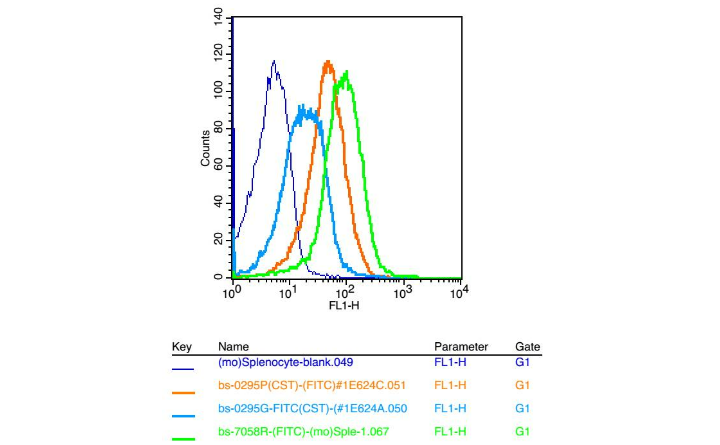
Positive control: mouse Splenocytes(2% Paraformaldehyde-fixed )
Isotype Control Antibody: Rabbit IgG Dilution: 1μg in 100 μl 1X PBS containing 0.5% BSA; Secondary Antibody: Goat anti-rabbit IgG-FITC; Dilution: 1:200 in 1 X PBS containing 0.5% BSA; Primary Antibody : rabbit Anti-EMR1 bs-7058R; Dilution: 1μg in 100 μl 1X PBS containing 0.5% BSA.
Isotype Control Antibody: Rabbit IgG Dilution: 1μg in 100 μl 1X PBS containing 0.5% BSA; Secondary Antibody: Goat anti-rabbit IgG-FITC; Dilution: 1:200 in 1 X PBS containing 0.5% BSA; Primary Antibody : rabbit Anti-EMR1 bs-7058R; Dilution: 1μg in 100 μl 1X PBS containing 0.5% BSA.

Blank control: Mouse brain.
Primary Antibody (green line): Rabbit Anti-EMR1 antibody (bs-7058R)
Dilution: 1μg /10^6 cells;
Isotype Control Antibody (orange line): Rabbit IgG .
Secondary Antibody : Goat anti-rabbit IgG-PE
Dilution: 1μg /test.
Protocol
The cells were fixed with 4% PFA (10min at room temperature)and then permeabilized with 90% ice-cold methanol for 20 min at-20℃. The cells were then incubated in 5%BSA to block non-specific protein-protein interactions for 30 min at at room temperature .Cells stained with Primary Antibody for 30 min at room temperature. The secondary antibody used for 40 min at room temperature. Acquisition of 20,000 events was performed.

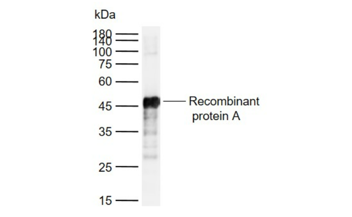
Sample:
U87MG(Human) Cell Lysate at 30 ug
Primary: Anti- EMR1 (bs-7058R) at 1/1000 dilution
Secondary: IRDye800CW Goat Anti-Rabbit IgG at 1/20000 dilution
Predicted band size: 95 kD
Observed band size: 95 kD
U87MG(Human) Cell Lysate at 30 ug
Primary: Anti- EMR1 (bs-7058R) at 1/1000 dilution
Secondary: IRDye800CW Goat Anti-Rabbit IgG at 1/20000 dilution
Predicted band size: 95 kD
Observed band size: 95 kD

Sample:
A549(Human) Cell Lysate at 30 ug
Primary: Anti- EMR1 (bs-7058R) at 1/1000 dilution
Secondary: IRDye800CW Goat Anti-Rabbit IgG at 1/20000 dilution
Predicted band size: 95 kD
Observed band size: 95 kD
A549(Human) Cell Lysate at 30 ug
Primary: Anti- EMR1 (bs-7058R) at 1/1000 dilution
Secondary: IRDye800CW Goat Anti-Rabbit IgG at 1/20000 dilution
Predicted band size: 95 kD
Observed band size: 95 kD

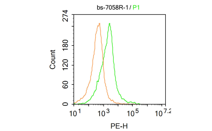
Blank control: Mouse kidney.
Primary Antibody (green line): Rabbit Anti-EMR1 antibody (bs-7058R)
Dilution: 1μg /10^6 cells;
Isotype Control Antibody (orange line): Rabbit IgG .
Secondary Antibody : Goat anti-rabbit IgG-PE
Dilution: 1μg /test.
Protocol
The cells were incubated in 5%BSA to block non-specific protein-protein interactions for 30 min at at room temperature .Cells stained with Primary Antibody for 30 min at room temperature. The secondary antibody used for 40 min at room temperature. Acquisition of 20,000 events was performed.

Sample:
A431(Human) Cell Lysate at 30 ug
Primary: Anti-EMR1 (bs-7058R) at 1/2000 dilution
Secondary: IRDye800CW Goat Anti-Rabbit IgG at 1/20000 dilution
Predicted band size: 95 kD
Observed band size: 95 kD
A431(Human) Cell Lysate at 30 ug
Primary: Anti-EMR1 (bs-7058R) at 1/2000 dilution
Secondary: IRDye800CW Goat Anti-Rabbit IgG at 1/20000 dilution
Predicted band size: 95 kD
Observed band size: 95 kD